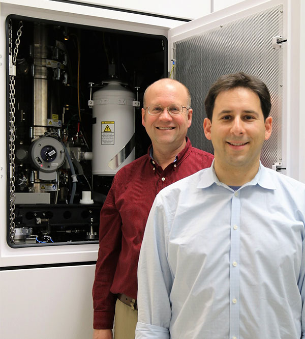
Sandia’s new state of the art transmission electron microscope
As Madonna once famously sang, we are living in a material world. Sandia now has a renewed ability to thrive in it, thanks to major recent upgrades in the Laboratories’ microscopy capabilities.
The centerpiece of a multiyear strategy to upgrade microscopy on Sandia’s California campus is a newly installed, state-of-the-art transmission electron microscope (TEM). The new instrument replaces two older instruments Sandia installed more than two decades ago, and brings the Labs powerful new capabilities for nanoscale analysis of materials.
The TEM works by sending a beam of electrons through a very thin material specimen and onto different sensors to form images or to measure composition. The sensitivity of the sensors in this TEM is higher than in the older machines. In addition, scientists have more control over the electron beam with this TEM.
The combination of increased sensor sensitivity and greater ability to control the beam mean that the TEM will not cook delicate organic or biological materials that usually become damaged under high-energy electron beams.
Materials scientist Josh Sugar says the high-speed, high-sensitivity sensor was developed for the detection of light in outer space and individual, sub-atomic charged particles. As it turns out, the sensor is extraordinarily good at detecting electrons as well. It can even measure a single, individual electron, which was previously not possible.
Another nice feature of this machine, Josh says, is that it offers higher energy resolution, which means researchers can learn more about the subtle details of the way atoms are bonded in a material.
Materials scientist Doug Medlin marvels at the precision and resolution of the new TEM compared to machines he used decades ago that required large photo plates. “When we would collect these types of images back in the day, we’d push the exposure button and we’d stop breathing, and wait until it did its thing,” Doug recalls. “Any motion in the room would blur the image.”
As Doug points out, Sandia has been a pioneer in atomic-scale materials simulations since the 1980s. Such simulations are important for predicting how the arrangement of atoms in a material can affect their mechanical or electronic properties. The capabilities of the new instrument provide a sensitive tool for testing how well these simulations actually work.
Not only can the TEM show the atoms that make up a material, it can also determine which elements those atoms are, and probe the 3-D microstructure of the material — all while people are walking around and talking in the control room. “When you have a new tool like this, you can make new discoveries,” Doug says.

Sandia materials scientists Doug Medlin (left) and Josh Sugar (right) pose in front of Sandia’s new transmission electron microscope. Even though the machine occupies most of the room, the samples it analyzes are no longer than a grain of rice and 1,000 times thinner than a sheet of paper.
State-of-the-art facility for a state-of-the-art instrument
This isn’t to say conditions don’t need to be controlled in the area surrounding the TEM. “For a high-end microscope like this you have to have high-end facilities: good control of air flow, magnetic fields, vibration, and temperature for it to reach its performance potential,” Doug says. “Otherwise it can affect your results.”
To illustrate how precisely the TEM’s environment is controlled, Doug reports that the temperature in the microscope lab fluctuates less than a degree Celsius over 24 hours. Furthermore, the electrical wiring and materials in the room and adjacent lab spaces were carefully considered to ensure the space would meet the stringent magnetic field requirements for the instrument. A lot of careful planning went into the creation of these conditions — all the more impressive given the short time frame in which the plans had to be executed.
Using the microscope facility as a pilot project for Sandia’s new integrated service delivery (ISD) approach, multiple groups on campus partnered to complete the project in eight months, start-to-finish.
“The ISD approach helped our cross-functional team to creatively plan, acquire the hardware, design and make significant laboratory modifications, as well as install and commission the microscope in about half the time it might otherwise have taken,” says Strategic Site Planning Manager Devon Powers.
Another important feature is that the microscope can be operated remotely, which will allow others to access the instrument directly from Albuquerque. Doug and Josh pioneered such remote microscopy several years ago so they could easily access an instrument based in New Mexico.
The result of all these features makes the TEM, along with the broader suite of Sandia’s microscopy tools and staff, a perfect complement for a wide range of scientific and engineering projects across the Laboratories. The following are just a few of the projects that will benefit.
Observing bubble behaviors and molecular “tinker toys”
The average person might think of helium as the gas that enables a fun voice-changing gag when inhaled from balloons at parties. But according to Josh, “helium is a specific scientific problem that really needs the new microscope.”
The problem is that nuclear weapons use tritium, a radioactive gas. Over time the gas can permeate into steel and decay, producing nanoscale helium bubbles that can crack critical structural components.
Josh has been working on imaging the bubbles inside the metal to inform behavior models. “The new TEM will make the imaging much easier, and that will help us design these systems, change manufacturing procedures, and make better predictions about how these things will work years down the road,” says energy nanomaterials group manager Andy Vance.
Similarly, research on a new class of materials called metal organic frameworks (MOF) also stands to benefit from the TEM. MOFs are crystalline materials in which metal ions are linked to organic (carbon-containing) molecules to create a structure. Their regularly spaced pore channels can be modified to accommodate guest molecules of various sizes, and they sport extremely high surface areas.
These properties make them applicable to a dizzying array of applications, including removing volatile radioactive gas from spent nuclear fuel, chemical and radiation detection, microelectronics, hydrogen storage for fuel cell vehicles, and even breathalyzers that indicate infectious diseases.
Chemist Mark Allendorf has been studying MOFs for more than a decade, and refers to them as “tinker toys” for chemists because like the children’s toy, the material constituents can be assembled like molecular scaffolding.
Previously, however, it was not possible to look at them using a traditional electron microscope because MOFs are very beam sensitive. The TEM’s direct-electron sensor will allow Mark and other scientists to look at the atomic structure of MOFs and at defects in their structure, which can affect properties such as electrical conductivity and gas storage.
A big suite for tiny things
The tools in Sandia’s microscopy suite complement each other, offering scientists a robust set of tools that can help them link what they see on many different length-scales. For example, another machine on site, the focused ion beam scanning electron microscope (FIB-SEM) can perform multiple functions that complement the new TEM, but at a larger-length scale than the TEM (from millimeters to tens of nanometers). It can also fabricate specimens appropriate for atomic-resolution analysis in the TEM.
An SEM uses similar operating principles, but instead of looking through a sample the way a TEM does, the electrons bounce off the surface of a material and generate a detailed image of its topography. It’s more like a high-powered pair of binoculars compared to the TEM, where the electrons go through a sample and you get something like a medical X-ray image. The California campus is planning to install a new SEM this year as well.
Other recent microscopy upgrades include new polishing equipment in the metallography laboratory where materials are prepared for the microscopes, new arrangement of lab space that make it easier to work in them, and new additions to the microscopy team, like Alan Jankowski, who were hired to facilitate the increased flow of work that the new tools will likely generate.
According to Josh, nothing less than the future of mankind could be determined by the materials that we make. “When I think about the history of civilization and man, I think about the materials that were used. There’s the stone age, the bronze age, the iron age. In the stone age, we lived in caves and we had stone tools and we hunted,” he says. “Very much, the materials that society used dictated what technologies were available to us and how we lived. And every time we had a major change in the materials that we used, our society and civilization had a major change in what we could do.”
Historically, advances in materials have been enabled by parallel advances in technologies to investigate them. These recent investments in materials characterization will help Sandia continue to be a major force in this field.